Research
|

|
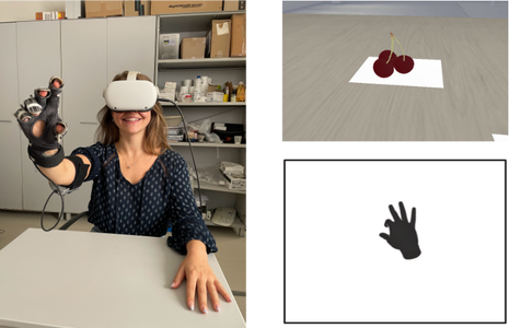
Rewiring the evolution of the human hand: How the embodiment of a virtual bionic tool improves behaviorMarucci M., Maddaluno O., Ryan C.P., Perciballi C., Vasta S., Ciotti S., Moscatelli A. & Betti V. (2024) iScience
|

|
|
|
|

|
|
|
Grounded Cognition: nuove prospettiveMarucci M., Betti V. (2021) Reti, saperi, linguaggi
|

|
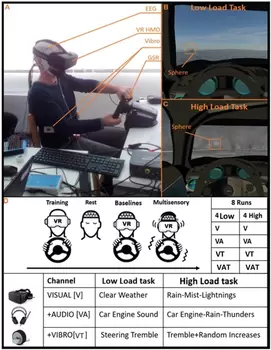
The impact of multisensory integration and perceptual load in virtual reality settings on performance, workload and presenceMarucci M, Di Flumeri G, Borghini G, Sciaraffa N, Scandola M, Pavone EF, Babiloni F, Arico P., Betti V (2021) Scientific Reports
|
|
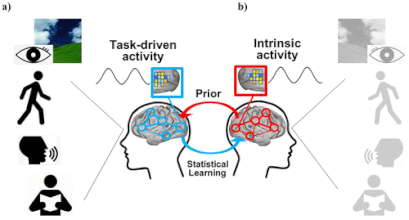
Spontaneous beta band rhythms in the predictive coding of natural stimuliBetti V, Della Penna S, de Pasquale F, Corbetta M (2021) Neuroscientist |
|
|
Leone F., Caporali A., Pascarella A., Perciballi C., Maddaluno O., Basti A., Belardinelli P., Marzetti L., Di Lorenzo G., Betti V.
|
Valutazione e trattamento dell’equilibrio nell’invecchiamento e nel paziente neurologicoMarco Tramontano, Simona Vasta , Gabriele Marangon , Amaranta Soledad Orejel Bustos , Viviana Betti
|
|
EXAMINING SELF-GENERATED ACTION RECOGNITION THROUGH
|
|
HIGH-DENSITY ELECTROENCEPHALOGRAPHIC CORRELATES OF GAIT DISORDERS IN POST-STROKE PATIENTSG. Marangon, S. Vasta, A. Orejel Bustos, R. Montemurro, V. Betti, M. Tramontano
|
Entropia: un universo di connessioni tra fisica, vita quotidiana e neuroscienzeVasta S., Marangon G., Orejel Bustos A.S., Tramontano M., Betti V.
|
ENCODING DEXTERITY IN THE HUMAN BRAIN |
|
Spectral connectivity fingerprint in the resting human brain: a MEG study
Maddaluno O., Guidotti R., Vettoruzzo A., Marzetti L., Cisotto G., Betti V.
|
|
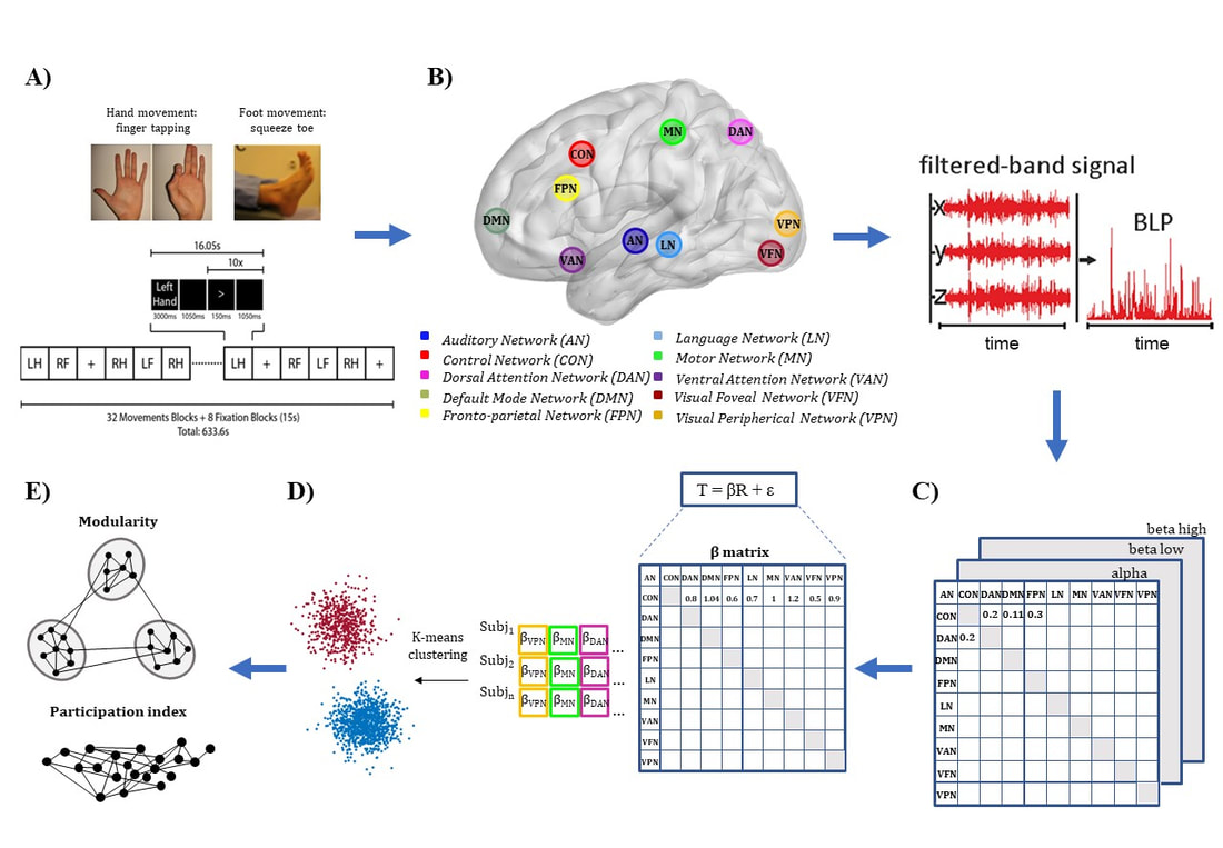
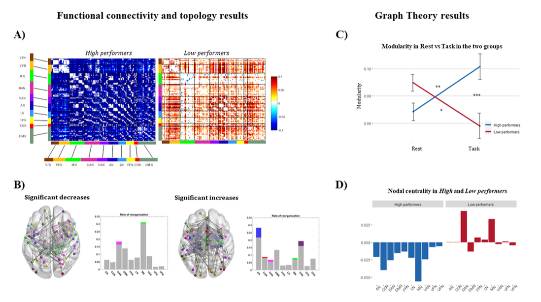
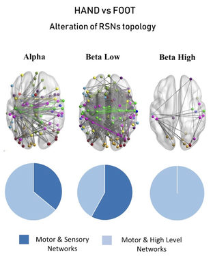
Distinct MEG functional connectivity and topology changes distinguish between voluntary hand and foot movementsPizzuti A, Della Penna S, Spezialetti M, Corbetta M, Betti V
|
|
On the contribution of visual statistics to changes of MEG functional connectivity patternsChiara De Giorgi , Sara Petrichella , Paolo Papale , Andrea Leo , Emilano Ricciardi , Pietro Pietrini , Maurizio
Corbetta , Stefania Della Penna , Viviana Betti |
|
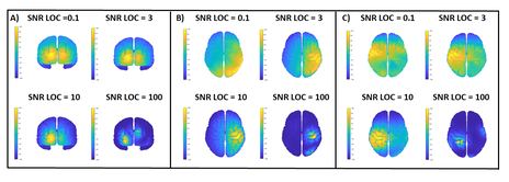
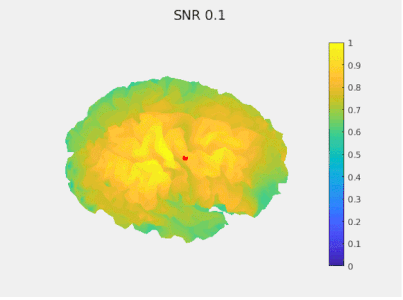
How Signal-to-Noise Ratio affects source reconstruction with Minimum Norm: the case of high-density EEG resting statePizzuti A., Basti A., Maddaluno O., Belardinelli P., Marzetti L., Di Lorenzo G., Betti V.
Awarded as best oral presentation |
|
Representation of hand shape in the human resting-state activityEl Rassi Y., Handjaras G., Leo A., Papale P., Corbetta M., Ricciardi E., Betti V.
|
|
papers IN preparation
Stability and flexibility of the intrinsic network connectivity associated with manual dexterity: a MEG study
Representation of the hand in human resting-state activity
States and transitions of everyday manual behavior
On the contribution of visual statistics to changes of MEG functional connectivity patterns
Follow us on social networks:
